Late autumn through winter is a premium time to encourage clients concerned by dark spots and other skin discolourations to seek solutions for one of Australia’s biggest skin concerns – hyperpigmentation.
Treatments are best performed during these months as clients are typically less exposed to UV on a daily basis; spending less time outdoors and wearing more “covered” clothing.
Dora Erdossy* is a leading industry educator on the protocols and products for the treatment of various forms hyperpigmentation.
But to treat clients effectively, you need to know the source of a client’s pigmentation.
Dora explains the various causes as we head into the peak season for offering treatments:
As skin therapists and dermal clinicians, there are a number of skin conditions we are presented with on a daily basis and sometimes providing appropriate solutions and positive outcomes for these conditions are not easy to come by.
There is one skin condition for clients that is visibly concerning, as they hope for for results as soon as possible but continues to be one of the most challenging one for us as professionals to treat – hyperpigmentation.
Melanogenesis – A Brief Overview
Our melanin-producing cells, melanocytes, are found in the stratum germinativum and form part of the epidermal melanin unit which includes keratinocytes cells. Melanogenesis occurs within melanosomes that are membrane-bound organelles and are the metabolic unit of the melanocyte cell.
The formation of pigment is highly complex but is initiated by the key enzyme tyrosinase.

Tyrosinase is located in the membrane of melanosomes and is responsible for the conversion of dopa to melanin. The melanin pathway is divided following the formation of dopaquinone into the production of eumelanin (brown-black pigment) and pheomelanin (red-yellow pigment). Both are found in all skins but in varying amounts.
Melanocytes are dendritic cells: that is, they have spidery-like projections that extend in all directions from the melanocyte body allowing melanosomes to be transferred to the keratinocytes via these dendrites.
As keratinocytes move towards the surface of the skin they carry the pigment with them, giving the colour we see on skin.
Interestingly, there are no racial differences in the overall number of melanocytes. However, it is the number and size of melanosomes that are different; for example, darker skin types have larger melanosomes.
Skin Pigmentation Triggers
There are certain external and internal factors that can affect the production of melanin and it is important to be aware of these to ascertain why your client is perhaps experiencing what they are. Let’s look at some common triggers:
Sun Exposure
As beautiful and health-giving as the sun is, it is the number 1 cause of hyperpigmentation. Sun exposure directly triggers the production of melanin and enhances the effect of any other trigger.
It is also ultimately responsible for the appearance of all types of dark spots that are created by melanin.
Sun exposure also darkens the colour of existing spots such as freckles and post- inflammatory hyperpigmentation (PIH).
Inflammatory Processes
Inflammation of the skin can occur when the skin has been put under stress due to certain situations such as in-clinic treatments including laser treatments and chemical peels or trauma to skin as seen in acne skin.
Melanocytes respond to the skin insult by causing an increase in melanin production and consequently darkening the area of skin injury creating PIH, which may prove difficult to treat. This is prevalent mostly in higher Fitzpatrick skin types.
Photo-Sensitising Medications
There are certain medications such as antihistamines, antidepressants and antibiotics that can cause photosensitivity.
The skin responds due to the interaction between the photo-sensitising chemical delivered to the body and exposure to either sunlight or a source of artificial radiation. As a result, irregular patches of hyperpigmentation appear in sun exposed areas.
Hormonal Changes
Melasma is the most common hyperpigmentation disorder due to hormonal changes.
Exacerbating factors include pregnancy and hormonal therapy such as oral contraceptives or Hormonal Replacement Therapy (HRT) for menopause.
Throw sun exposure into the equation and melasma is made worse!
How to effectively analyse hyperpigmentation
Hyperpigmentation can be a tricky skin condition to analyse effectively just with the naked eye so l often recommend the use of a Woods Lamp or an equivalent device to be able to classify what type of pigmentation you are treating.
This enables us to determine what we realistically can treat in relation to the skin care and modalities we have on offer:
Epidermal Pigmentation
This form of pigmentation is a dark brown colour with very obvious demarcated borders. There is an excessive amount of melanin in all epidermal layers but with a normal amount of melanocytes.
Dermal Pigmentation
These lesions are not as intensified and are of a brown-grey colour with not very obvious borders. Melanin is found between collagen bundles or within melanophages, which are macrophages that have engulfed melanosomes.
Mixed Pigmentation
When examining with a Woods Lamp or other skin analysis device, pigmentation appears to be more obvious in some areas and not so obvious in others. It is characterised by an increase in epidermal melanin and many melanophages in the dermis. The epidermal component is intensified the most.
Examples of Hyperpigmentation Disorders
Ephelides
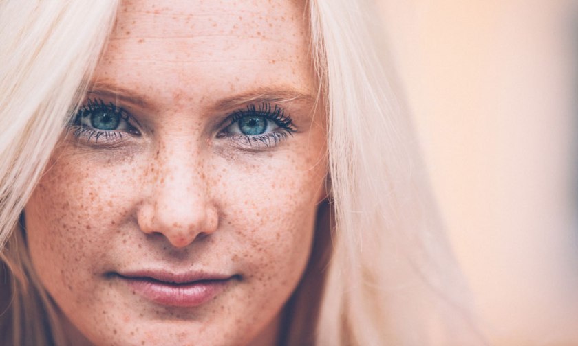
Ephelides (freckles) are commonly seen in fair-skinned individuals and are the result of an increase in UV-induced melanogenesis as well as an increase in the number of melanosomes transferred from melanocytes to keratinocytes.
They appear on sun-exposed areas such as the face and the back of the hands and are well-demarcated, hyperpigmented macules that are generally 1-3mm in size.
Lentigines
Solar lentigines are also commonly seen in fair-skinned individuals aged over 60 on sun-exposed areas, especially the face, back, forearms, backs of hands and the upper chest. The prevalence of solar lentigines increases as one gets older. They appear as macules or patches that are either oval, round or irregularly shaped. They can vary in colour from tan to dark brown and can range from 3mm to 2cm in size.
Post Inflammatory Hyperpigmentation (PIH)
As mentioned, PIH is commonly seen in higher Fitzpatrick skin types and is a consequence of the melanocytes response to skin injury. Post-inflammatory hyperpigmentation can appear as small to large macules and/or patches.
Melasma
Melasma appears as a number of hyperpigmented patches that range from either one lesion to many patches and are usually located symmetrically on the face.
Three clinical patterns of melasma are identified based on the distribution of lesions:
- Centrofacial pattern: This is the most common pattern and it involves the forehead, cheeks, upper lip, chin and nose
- Malar pattern: The nose and cheeks are involved
- Mandibular pattern: This pattern involves the ramus of the mandible (jaw)
So what can be done?
Treating hyperpigmentation is an ongoing challenge for us as skin professionals but nevertheless we do have therapies which could help make a difference, that range from topical solutions to physical modalities.
First and foremost what l ALWAYS discuss with PASSION is the importance of a thorough and detailed consultation to ascertain what the potential trigger is but also determine how realistic an outcome we can provide for the client.
Educating the client is also key to ensure that they are contributing to a positive outcome such as avoiding sun exposure, which is the primary cause of hyperpigmentation.
The importance of using a broad spectrum sun protection must also be stressed to not only assist in the prevention of hyperpigmentation but also reduce the incidence of PIH, especially if we are choosing to perform certain treatments that could increase the risk of PIH such as light based or laser therapies or even chemical peels.
If the client does not adhere to this advice, then the failing of the chosen treatment regimen is inevitable.
In saying this, treating pigmentation disorders during winter maybe a viable option especially for those clients who may not be so good in following your advice in avoiding the sun.
Along with laser and light therapies and chemical peels, there are unique methods such as the Cosmelan Method, which can assist in effectively treating hyperpigmentation disorders.
This incorporates an in-clinic treatment as well as strict homecare and has proven to provide fantastic results for hyperpigmentation disorders including melasma which, as we know, is a challenging disorder to treat.
The use of topical ingredients such as kojic acid, niacinamide (vitamin B3), ascorbic acid, N-Acetylglucosamine and retinol (vitamin A derivative) can help to disrupt the pathways of melanogenesis.
Incorporating topical ingredients, hence homecare for hyperpigmentation, is highly recommended in combination with in-clinic treatments to help maximise results.
In conclusion …
The formation of melanin is a highly complex process and is involved in many processes such as sun exposure and inflammation.
Hyperpigmentation disorders such as melasma and PIH are not only skin conditions that are visibly concerning to clients but conditions that clients hope to achieve positive outcomes for as soon as possible.
As skin therapists and dermal clinicians, not only should we be fully knowledgeable of melanogenesis and the many triggers which can overstimulate this process but we must also be knowledgeable of the product and treatment solutions we have on offer and understand what we can realistically achieve for the client.
Providing an outcome that is both positive and realistic for the client and us can be difficult when treating hyperpigmentation. However if we educate the client accordingly of what is possible as well as what they should be doing to assist in the process.
The end result can potentially be a happy client and isn’t that ultimately what we are always striving for in what we do on a daily basis …making our clients happy?!
ABOUT DORA ERDOSSY AND COSMELAN
Dora is the national trainer (east coast Australia) for Cosmelan products and protocols, distributed by Advanced Cosmeceuticals, and is currently lecturing for the Dermal Science degree at Victoria University.
Cosmelan is produced by Mesoestetic, a leading specialist in cosmeceutical skincare, and is an effective topical skin brightening treatment that helps to visibly lighten dark spots and blemishes caused by excess melanin.
The Cosmelan Treatment features a highly effective formula that helps interrupt the main steps in the melanin synthesis pathway. It is a two-stage process:
Stage 1: Professional treatment in the salon or clinic
Stage 2: Home-care treatment pack.
During treatment, it is important to protect the skin at all times using an ultra-high SPF sunscreen, reapplying regularly throughout the day. This will ensure that the treatment is effective as it helps prevent hyperpigmentation.
Results
After the second or third week of Cosmelan treatment, the appearance of the skin is visibly improved. Dark spots are lighter and less noticeable while the skin has a more youthful appearance and radiant glow overall.
It has shown an effectiveness rate of 95-99% of cases of skin spots and hyperpigmentation tested.
In 2015, Mesoestetic launched an international campaign to develop a Specialised Depigmentation Centre program.
The program is designed to qualify and certify an exclusive, select number of high quality skin care clinics as specialised depigmentation centres, with 42 clinics across Australia participating in the launch global initiative.
Pigmentation is perceived as the third most important skin problem after wrinkles and sagging.
More than 90 percent of Caucasian people aged over 50 have skin blemishes. Today, depigmenting treatments represent over 20 percent of the total cosmetic market.
“We are very proud to have had the opportunity to offer this initiative to our qualifying clients to become Mesoestetic Depigmentation Specialist Centres,” says Catherine Biedermann, managing director of Advanced Cosmeceuticals.
“Not only has this course provided them with a competitive advantage but they are also part of a global skincare community that Mesoestetic is establishing.”
The training covers the importance of taking into consideration a client’s commitment to treatment.
For instance, the Cosmelan treatment requires a 4-month commitment and if the client is unable to commit, a different treatment should be recommended that would be more suitable.
Ongoing training and support is an integral part of this exclusive scheme, with qualifying clinics committed to attending training schools and in-clinic training.
In order to be able to do this effectively and with confidence, the clinician must have comprehensive knowledge of the product range, composition of the products at hand, how to use them with machines, protocols of application and home care recommendations – making continuous training an absolute must.
REFERENCES
Gillbro, J.M, & Olsson, M.J. (2011). The melanogenesis and mechanisms of skin-lightening agents – existing and new approaches. International Journal of Cosmetic Science, 33, 210-221.
Nicolaidou, E & Katsambas, A.D. (2014). Pigmentation disorders: hyperpigmentation and hypopigmentation. Clinics in Dermatology, 32, 66-72.
Nieuweboer-Krobotova, L. (2012). Hyperpigmentation: types, diagnostics and targeted treatment options. Journal of the European Academy of Dermatology and Venereology, 27(1), 2-4.
Vashi, N.A., & Kundu, R.V. (2013). Facial hyperpigmentation: causes and treatment. British Journal of Dermatology, 169(3), 41-56
REFERENCES
Gillbro, J.M, & Olsson, M.J. (2011). The melanogenesis and mechanisms of skin-lightening agents – existing and new approaches. International Journal of Cosmetic Science, 33, 210-221.
Nicolaidou, E & Katsambas, A.D. (2014). Pigmentation disorders: hyperpigmentation and hypopigmentation. Clinics in Dermatology, 32, 66-72.
Nieuweboer-Krobotova, L. (2012). Hyperpigmentation: types, diagnostics and targeted treatment options. Journal of the European Academy of Dermatology and Venereology, 27(1), 2-4.
Vashi, N.A., & Kundu, R.V. (2013). Facial hyperpigmentation: causes and treatment. British Journal of Dermatology, 169(3), 41-56.